What is the correct treatment for tarus (buckle ) fractures of distal radius in children

5 year old with injury to forearm due to fall in the playground
How do you manage these fractures?
a above elbow plaster
b below elbow plaster
c splints
How long will you continue immobilisation?
a 2 weeks
b 3 weeks
c 4 weeks
d 6 weeks
How often will you repeat the rays?
- every week
b After 3 weeks
- At week 1 and at plaster removal
Here is an attempt to find an answer to some of the questions…
What does the term tarus fractures mean
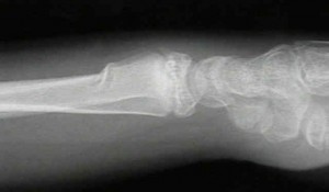
Torus is derived from Latin (tori) meaning a swelling or protuberance. Torus (buckle) fractures of distal metaphysis of radius & ulna are the most common fractures of in lower forearm in young children
When the child falls on the outstretched hand, the bone buckles under the weight of the body. Typically, you will see a slight bend in the bone, but only on one side. It literally looks like the bone buckled, but didn’t break all the way through.
There is usually little if any deformity in the bone because the periosteum and cortex are intact on the side. Technically speaking, the bone has failed in ‘compression’, making the small ripple in the bone that you see on the x-ray.
Torus fractures hurt, but will not cause a visible deformity. If you push directly on the arm where it is broken, it will hurt, and the child can localize the spot very clearly. However, they can usually move the wrist joint without much trouble. Because of this, many of these fractures are diagnosed late because it can be difficult for parents to tell if it is broken or not
These are generally stable fractures, meaning that the alignment will probably not change with protection such a a cast or splint.
What is the evidence from the literature
- Davidson (JBJS B 83-B 20011173-1175 ) did a RCT comparing treatment in traditional forearm cast V/S splint and concluded that there was no difference in the results of both the treatment methods. In fact the splint treatment had major benefits in terms of cost andreduction of the number of attendances.
- Pint et al (Pediatrics. 2006 Mar;117(3):691-7) in a radomised control trial of 87 fractures : 42 in the splint group and 45 in the cast group concluded that children treated with removable splinting have better physical functioning and less difficulty with activities than those treated with a cast. Splinted children had less difficulty with bathing throughout the entire study. There were no significant differences in pain between groups as measured by visual analog scale. There were no refractures.
- Farbman KS 9Arch Pediatr Adolesc Med September 1999;153:923-5.) while evaluating the role of serial radiographs in the management of pediatric torus fractures concluded that radiographs after application of a cast for a nondisplaced torus fracture are not required. Clinical examination is appropriate during follow-up, with consideration given to repeat radiographs only at four weeks postfracture to document healing.
What are the pitfalls
- Greenstick fractures of lower radius: need to be reduced and put in cast as they can deform and redisplace. The failure in greenstick fractures in distraction and is on the side of angulation
- Eren (Journal of Pediatric Orthopaedics B:January 2009 – Volume 18 – Issue 1 – pp 35-36) reported onTorus fracture of the distal radius associated with distal radioulnar joint instability. They suggested that torus fractures of the children are not always innocent, and simple physical examination of the DRUJ for tenderness and instability is mandatory before and after roentgenographic evaluation to adequately treat these kinds of injuries and prevent malpractice. In cases of doubt, MRI examination of the wrist may also reveal the torn triangular fibrocartilage complex leading to instability.
- Some of the buckle fractures may have extension to the physis . Aminian A, Schoenecker PL (J Pediatr Orthop. 1995 Jul-Aug;15(4):495-8) reported on premature closure of the distal radial physis after fracture of the distal radial metaphysis.
So coming back to the case how do you manage the 5 year old child with the tarus fracture of distal radius and ulna?
Will wait for the responses from the readers…
References
Simple treatment for torus fractures of the distal radius
- S. Davidson, FRCS Orth, Specialist Registrar in Orthopaedic Surgery; D. J. Brown, FRCS, Specialist Registrar in Orthopaedic Surgery; S. N. Barnes, FRCS, Specialist Registrar in Orthopaedic Surgery; and C. E. Bruce, FRCS Orth, Consultant in Paediatric Orthopaedics From Alder Hey Children’s Hospital, Eaton Road, West Derby, Liverpool L12 2AP, UK.
Journal of Bone and Joint Surgery – British Volume, Vol 83-B, Issue 8, 1173-1175.
doi: 10.1302/0301-620X.83B8.11451
Correspondence should be sent to Mr D. J. Brown at 6 Caldy Road, West Kirby, Wirral CH48 2HG, UK.
Torus (buckle) fractures of the distal radius are common inchildhood. Based on the results of a postal questionnaire anda prospective, randomised trial, we describe a simple treatmentfor this injury, which saves both time and money.
Over a six-month period, we randomised 201 consecutive patientswith this injury to treatment with either a traditional forearmplaster-of-Paris cast or a ‘Futura-type’ wrist splint.All patients were treated for a period of three weeks, followedby clinical and radiological review.
There was no difference in outcome between the two groups, andall patients had a good result. Only one patient did not toleratethe splint which was replaced by a cast.
The questionnaire showed a marked variation in the way in whichthese injuries are treated with regard to the method and periodof immobilisation, the number of follow-up visits and radiographstaken.
We suggest that a ‘Futura-type’ wrist splint canbe used to treat these fractures. The patient should be reviewedon the following day to confirm the diagnosis and to give appropriateadvice. There is no evidence that further follow-up is required.
This simple treatment has major benefits in terms of cost andreduction of the number of attendances.
A randomized, controlled trial of removable splinting versus casting for wrist buckle fractures in children.
Plint AC, Perry JJ, Correll R, Gaboury I, Lawton L.
Source
Department of Pediatrics, University of Ottawa, Ottawa, Ontario, Canada. plint@cheo.on.ca
Pediatrics. 2006 Mar;117(3):691-7.
Abstract
OBJECTIVE:
Wrist buckle fractures are a frequent reason for emergency department visits. Although textbooks recommend 2 to 4 weeks of immobilization in a short arm cast, management varies. Treatment with both casts and splints is common, and length of immobilization varies. The objective was to determine if children with distal radius and/or ulna buckle fractures treated with a removable splint have better physical functioning than those treated with a short arm cast for 3 weeks.
METHODS:
This was a randomized, controlled trial in the emergency department of an academic, tertiary care children’s hospital. Participants were children 6 to 15 years of age with distal radius and/or ulna buckle fractures who were randomly assigned to treatment with a short arm cast for 3 weeks or a removable splint. Cast removal was at 3 weeks. A validated self-reported outcome tool, the Activities Scales for Kids performance version (ASKp), was used to measure physical functioning over a 4-week period. The main outcome was the ASKp score at 14 days postinjury.
RESULTS:
We randomly assigned 113 patients, and 87 were included in the final analysis: 42 in the splint group and 45 in the cast group. Study groups were similar in age, gender, bone fractured, and dominant hand injured. There were significant differences in ASKp score at day 14 and change in ASKp from baseline at days 14 and 20, indicating better functioning in the splint group. Splinted children had less difficulty with bathing throughout the entire study. There were no significant differences in pain between groups as measured by visual analog scale. There were no refractures.
CONCLUSIONS:
Children treated with removable splinting have better physical functioning and less difficulty with activities than those treated with a cast.
Are Serial Radiographs Needed in Torus Fractures in Children?
March 17, 2010
American Family Physician, Feb 15, 2000 by Jeffrey T. Kirchner
A torus, or “buckle,” fracture of the distal radius is a common type of fracture in children. The standard treatment for these nondisplaced fractures is casting for three to four weeks. Serial radiographs are often obtained, such as immediately after application of the cast and again a week or so later. To determine whether repeat radiographs are necessary, Farbman and colleagues assessed the utility of follow-up radiographs in 70 children (46 boys and 24 girls) with torus fractures of the distal radius or ulna.The authors’ medical center had no standard protocol for follow-up radiography in patients with torus fractures. Radiographs before and immediately after casting were obtained in the emergency department in 24 of the children.
All but five of the 70 children were subsequently seen in the orthopedic clinic at least four weeks after the injury. Repeat radiographs were obtained as early as two days and as late as seven weeks after the fracture. Follow-up films were obtained twice in 21 children, three times in 12 children and four times in five children. The most common reasons for repeat radiographs were documentation of healing and diagnostic confirmation. The casts remained in place for at least two weeks in all of the children, and casts were removed before the fourth week in 20 children.
The average number of follow-up radiographs was 3.3 studies per patient. At a cost of $119 per study at the authors’ institution, this translates to a total cost of $27,251 for radiology services rendered to the 70 patients in the study. The authors cite a study of follow-up radiographs in adults with stable ankle fractures. That study revealed that an average of 4.5 ankle radiographs per patient was obtained, but no radiographs demonstrated a change in fibular alignment in any patient. The authors calculated that as much as $35 million would be saved annually if routine follow-up radiographs were not performed in patients with torus fractures.
To compare protocols for the management of torus fractures at other hospitals, the authors sent a survey to the directors of seven orthopedic surgery residency programs in the New England area. Responses were received from all seven directors. Each said that they did not advise their residents to obtain radiographs after application of a cast in patients with a torus fracture. However, one director noted that the emergency department at the hospital had a standing policy to do so. In response to the question of why repeat films may be obtained, one residency director noted medicolegal reasons, and another mentioned educational purposes, to assess proper placement of a cast.
The authors note that there are no national guidelines that stipulate any medical indications for postcasting radiographs in children with torus fractures, nor does the medical literature support the need for follow-up radiographs one or two weeks after the fracture. Repeat films are recommended to check for displacement after casting only in the presence of dorsal angulation and bicortical fracture or of unicortical fracture through the entire bone to the opposite cortex.
The authors conclude that radiographs after application of a cast for a nondisplaced torus fracture are not required. Clinical examination is appropriate during follow-up, with consideration given to repeat radiographs only at four weeks postfracture to document healing